Nenhum resultado de conteúdo corresponde à sua palavra-chave.
Resultados
Encerrou sessão com sucesso.
Ainda não está registrado?
Descubra novos espaços para a fusão
Inspiradas na anatomia humana e potenciadas pela ciência, nossos implantes combinam avanços tecnológicos com valores clínicos. O resultado é um grande avanço na estabilização anterior e posterior.
Structan®
Você acha que isso é uma rede de implante comum? Bem, impressione-se com a ciência por trás do Structan®. Décadas de experiência, combinadas com tecnologia moderna, levaram à sua criação – uma estrutura projetada para melhorar os resultados clínicos e o desempenho biomecânico avançado.
A área da superfície é ampliada por
0
vezes, proporcionando mais oportunidades para o crescimento ósseo.
Resistente e elástico ao mesmo tempo – Structan® é
0%
mais próximo do módulo de elasticidade do osso cortical. (1-4) *
Cubra a estabilização posterior apenas com
0
plataforma espinhal modular que se adapta com precisão às suas necessidades.
Dispositivos de fusão intercorporal 3D AESCULAP®
Assim como na criação de todas as nossas soluções para a coluna vertebral, o design dos dispositivos de fusão intercorporal AESCULAP® 3D baseia-se nos nossos valores fundamentais de criação de desempenho biomecânico avançado, flexibilidade intraoperatória e melhores resultados clínicos.
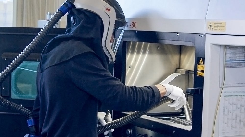
Fabricação aditiva

Structan®
O portfólio


Animações do fluxo de trabalho cirúrgico
Veja o desempenho dos dispositivos de fusão intercorporal 3D AESCULAP® e Ennovate®.
Nosso dispositivo de fusão intercorporal TLIF, com seu insersor articulado, permite procedimentos de fusão verdadeiramente minimamente invasivos.
Com sua técnica cirúrgica simplificada, nosso dispositivo de fusão intercorporal PLIF e a Ennovate® são ideais para a abordagem aberta.
Combinando a essência de dois mundos – nosso dispositivo de fusão intercorporal TLIF pode ser implantado de forma minimamente invasiva ou aberta.
Descubra a plataforma espinhal AESCULAP®
*compared to solid titanium alloy interbody fusion devices.


